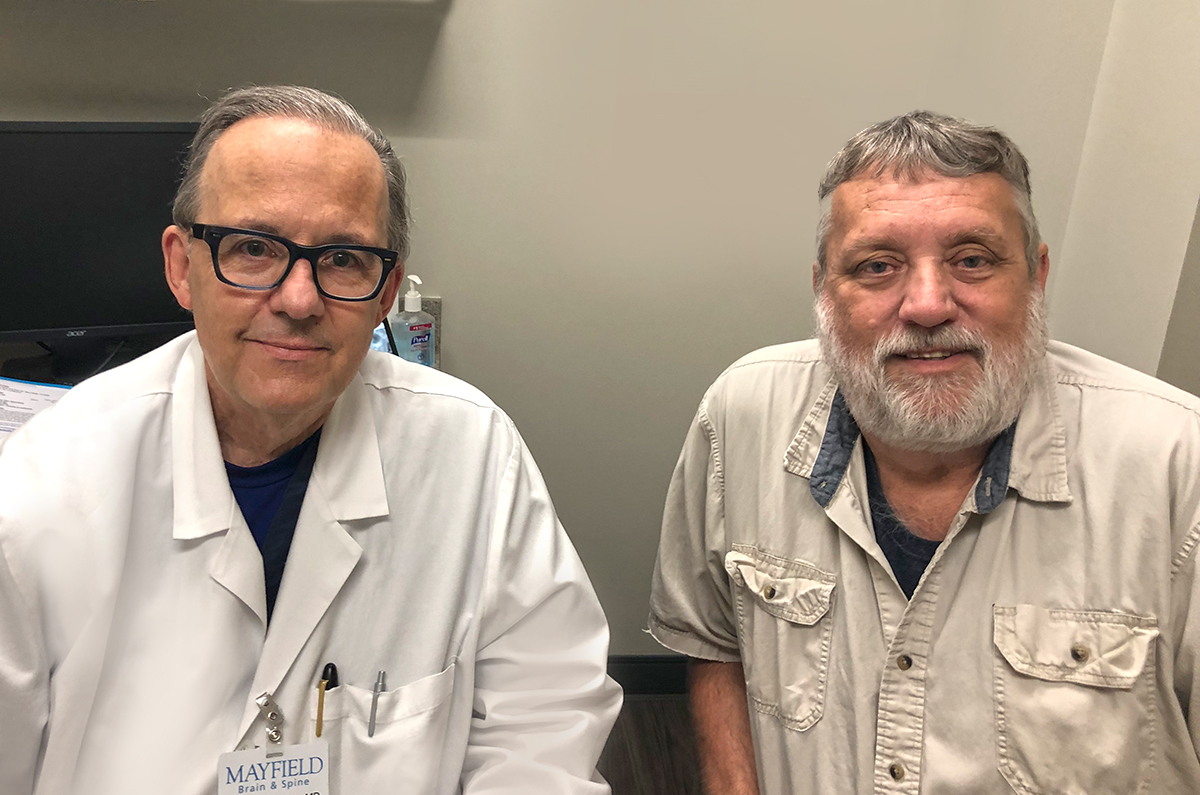
Mike's story
Pituitary adenoma
Second surgery, radiosurgery help Mike with recurrence of brain tumor
"I thought I was cured once already," Mike said.
Nearly 20 years after surgery to remove a tumor inside his skull, Mike started having symptoms again. He suffered from some migraine headaches, and his peripheral vision had narrowed significantly. A scan showed that fragments from the original tumor had recurred, or regrown, adjacent to his pituitary gland. Now slightly larger than a golf ball, it would have to be removed through another surgery.
"When the doctor called me and said, "Your tumor is back," I wasn't too worried," Mike said, "because I knew what to expect."
Mike was suffering from a pituitary adenoma, a tumor that grows from the pituitary gland, the bean-shaped organ that sits just behind the bridge of the nose, near the internal carotid arteries and the nerves that control eye movement. Pituitary tumors can occur in up to 15 percent of the population, and recurrence of the tumor after surgical removal is about as common. After his first surgery in 2001, Mike's medical team had monitored the tumor with regular scans for a decade, then reducing in frequency as the years passed.

With the most recent symptoms, fragments of the original tumor had grown into critically important areas that are hard to reach, around the right internal carotid artery and dangerously close to the optic nerve. Mike's primary care physician recommended consulting a brain surgeon, so Mike called Mayfield Brain & Spine, the same group that performed his first surgery 20 years before. This time, he was referred to neurosurgeon Dr. Vincent DiNapoli, who removed the tumor in a procedure at The Jewish Hospital lasting slightly longer than two hours. Dr. DiNapoli said brain tumors like the one that regrew for Mike can be challenging to remove completely because they sometimes are located adjacent to arteries, nerves, bone or other structures.
"We always continue to monitor patients for potential regrowth or recurrence," Dr. DiNapoli said. "Mike's symptoms were affecting his vision and his ability to work, and we felt the surgery could successfully remove the tumor and improve his daily life."
That May 2021 surgery wasn't the end of Mike's treatment. Fragments of the tumor had grown into hard-to-reach areas that are within a few millimeters of the optic nerve. The risk of further damage led Dr. DiNapoli to recommend Gamma Knife radiosurgery for Mike to eliminate the remaining tumor fragments, referring him to Mayfield neurosurgeon Dr. Ronald Warnick, a recognized radiosurgery expert.

Gamma Knife radiosurgery uses 192 beams of radiation and is often used as a less-invasive treatment option for brain tumors. Because the tumor fragments had grown so close to the optic nerve and could soon affect his vision, Dr. Warnick proceeded with Gamma Knife treatment, performed as an outpatient procedure in a single day, in the fall of 2021. He told Mike that his headaches should subside over time.
"Developing a layered treatment approach for Mike was the best way to address his brain tumor, minimize adverse effects and give him the best chance of a full recovery," Dr. Warnick said. "Once we identified that there was tumor remaining around the carotid artery within 2mm of the optic nerve, we wanted to intervene early to prevent growth that would lead to loss of vision. Gamma Knife radiosurgery provided the best option for ensuring his long-term outlook."
Today, Mike's headaches are gone, his overall health has improved, and he is positive about his outlook.
"I feel pretty good," he said. "I have confidence in Dr. DiNapoli, Dr. Warnick and the team, because I know they will watch my progress and monitor the tumor. Because of them, I've been able to deal with it."
~ Cliff Peale
Hope Story Disclaimer -"Mike's Story" is about one patient's health-care experience. Please bear in mind that because every patient is unique, individual patients may respond to treatment in different ways. Results are influenced by many factors and may vary from patient to patient.
