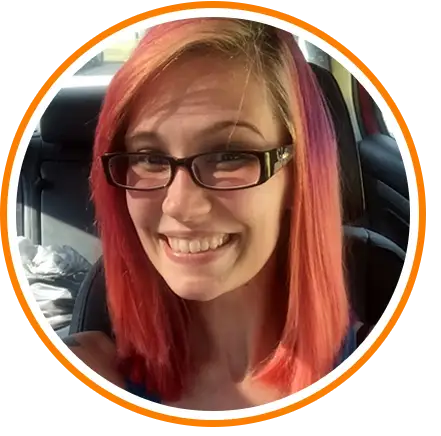
Jordin's story
Glioma brain tumor
One might say it was the storm before the calm. By the time Jordin had driven herself to Mercy Health – West Hospital, her head ached so badly that she could barely open her eyes. No, she did not have a history of headaches, she told the emergency room physician. No, the headaches during the last few weeks were not like anything she had ever experienced before.
A CT scan was immediately ordered, and by the time the results arrived, family members were there to hear the news: a lesion on the brain. An MRI with contrast followed, and the lesion came into clearer focus: a large tumor on the right parietal lobe of her brain.
"Of course we were all stricken with panic, thinking the absolute worst," Jordin recalls. "We were in shock. A nurse came back and said we needed to be seen immediately by a neurosurgeon."
Then came the calm.
The next morning Jordin and her family met with Vincent DiNapoli, MD, PhD, a neurosurgeon and brain tumor specialist with Mayfield Brain & Spine. Dr. DiNapoli spoke reassuringly with them and pulled up the images of Jordin’s tumor on a computer screen. He then scrolled through the images, slices of Jordin’s brain that unfolded millimeter by millimeter like an old-fashioned flipbook. Jordin, who had never seen an MRI before, was stunned.
"They start with your eyes and go to the back of your head," Jordin says. "And all of a sudden there’s this big blob in my brain. It glowed real bright. It was very scary. It was neat and terrifying at the same time."
Jordin's mother asked the question: "Is it cancer?"
"I’m 99 percent certain that it is not," Dr. DiNapoli replied. "The tumor does not light up with the contrast they gave, strongly suggesting that it is not malignant. It is likely slow-growing and has probably been there for some time."
"Dr. DiNapoli was super calm and put us at ease when we met him," Jordin says. "He explained everything, answered all of our questions. With the cancer part being set aside, we talked about what to do next."
Jordin’s options were to undergo radiation to stop the tumor from growing, or to undergo brain surgery and have it removed. "Almost in unison we said surgery," Jordin says. "We didn’t even really talk about it. I wanted the tumor out of my head."
Jordin's procedure took place at the University of Cincinnati Medical Center, where Dr. DiNapoli used advanced imaging and stereotactic navigation technologies to map a safe path to the tumor. The mapping included new, up-to-date MRI images taken just prior to surgery.
"That was probably the scariest day of my life," Jordin recalls. "I told Dr. DiNapoli that I was really scared. I was lying there crying. I hadn’t had anesthesia since I had my tonsils out in the third grade. He said, ‘Don’t worry, I’ll take good care of you. I do this every day.’ That made it a hundred times easier."
Over a period of five hours, Dr. DiNapoli and his surgical team removed all of the firm, grayish tumor, taking extreme care to remove all residual disease from the tumor’s border.
Complexities included the location of the tumor, which was within the motor planning area of the brain and, at its depth, adjacent to the visual pathways.
Later, the pathologist confirmed Dr. DiNapoli’s expectation that the tumor was not aggressive. It was a grade 2 glioma, the lowest-grade possible for an adult.
"It was the best we could hope for," Jordin says. "He got all of the tumor out, which is amazing. I’m so thankful for that. I spent three days in the UC brain tumor unit afterward and I had wonderful nurses. My boyfriend never left my side until it was time to go home. I was so thankful that I had them to take care of me."
Jordin experienced temporary loss of peripheral vision in her left eye following surgery, but her vision gradually returned and is now close to normal. Every now and then she experiences a mild headache, which she treats with Tylenol. Two months after her surgery she was cleared to return to work.
In the end, the young woman whose first reaction to her tumor was panic and shock began to take a calm, scientific interest in it. She even asked Dr. DiNapoli whether she could keep the tumor. A battle scar in a jar. He smiled and said no, but then posed an alternative: "You could donate it to science, and that way it might help someone else."
Jordin did. She donated her tumor to the University of Cincinnati’s tissue bank and also to the Ohio Brain Tumor Study. Jordin wanted to help others because her friends and family had done so much to help her through, and because a fellow patient had come forward to support her.
"We posted on Facebook what I was going through, and a friend from high school contacted me about his girlfriend, who had had the same thing, only cancerous, on her frontal lobe," Jordin says. "She helped me so much going through this. She gave me great advice: take it one day at a time.
"And that’s the advice I would give someone else who has been diagnosed with a brain tumor," Jordin continues. "It’s OK to have emotion. If you feel like crying, just cry. And it’s OK to be selfish. You are dealing with something that could be your life. Take one day at a time. It’s OK to keep pushing forward. Don’t let it get you; you have to get it."
Hope Story Disclaimer -"Jordin's Story" is about one patient's health-care experience. Please bear in mind that because every patient is unique, individual patients may respond to treatment in different ways. Results are influenced by many factors and may vary from patient to patient.


