Barbie's story
Meningioma/Craniotomy
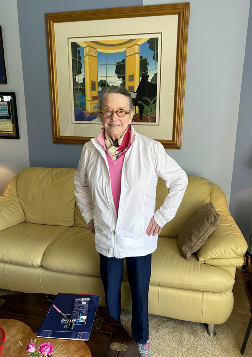
After brain tumor surgery, Barbie says, "I'm back!"
Barbara spent a professional lifetime as a writer, and her observation and wordsmithing skills are important to her. When she started to get more tired and her focus started to drift, it was a big deal.
"I was very fatigued, and I don't think I was quite as organized as I once was," recalls Barbara, whose friends call her Barbie. "I would just say to people, 'What's wrong with me?''
The symptoms had taken a huge toll on Barbie's daily life. She was spending most of her day in bed. She moved out of her house and in with a friend who has acted as her caregiver. She gave up driving, robbing her of her treasured independence. One doctor even raised the prospect that she might be suffering early signs of dementia.

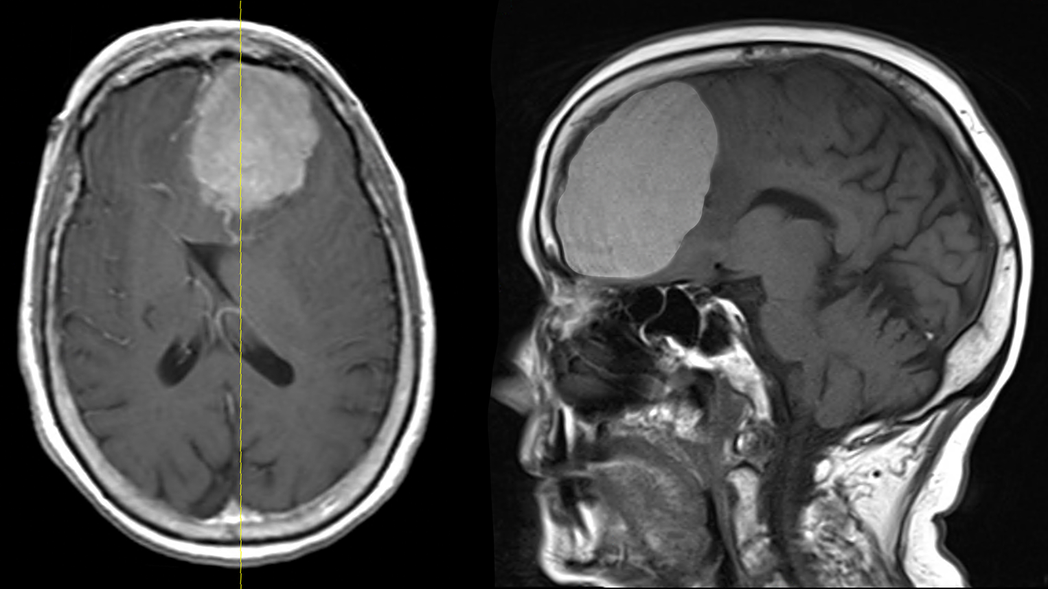
In early February, the answer came into focus when Barbie had trouble even waking up one day. Her friend took her to the emergency room, where doctors discovered a large benign tumor, known as a meningioma, lodged between the two hemispheres of her brain.
Meningioma tumors are slow-growing masses that originate in the covering of the brain, known as the meninges. They are the most common form of benign tumor. But this particular meningioma had grown to about the size of a baseball, causing significant pressure and swelling in the frontal lobes. These are the structures than govern basic functions like speech, energy and balance.
Barbie was referred to Dr. Brad Curt, a neurosurgeon at Mayfield Brain & Spine. Dr. Curt says the tumor had probably been growing for several years, but it was only after a gradual increase in symptoms became severe that it was discovered.
"The symptoms that she was noticing often can be attributed to other causes, and meningiomas grow slowly," Dr. Curt says. "The brain can accommodate that growth for a significant period of time. Once the tumor gets large enough, symptoms can worsen quickly, making it imperative to remove the tumor and relieve the pressure on the brain."
Dr. Curt was joined in the surgery by fellow neurosurgeon Dr. Yair Gozal. They performed a left frontal craniotomy, a common procedure where surgeons open the skull to gain access to the tumor. They were able to remove the entire mass, allowing the brain's frontal lobes to begin recovering.

"The parafalcine meningioma was located between the two hemispheres of the brain," Dr. Gozal says. "Tumors of this size compress brain tissue and can affect behavior and personality, but they often are difficult to diagnose because the symptoms increase gradually. And once the tumor is removed, it can take a substantial amount of time for the swelling to reduce and brain to fully recover."
Physical therapy helped Barbie regain her movement and alleviated her risk of falls. Six weeks after the surgery, her speech had improved and she was moving around with a walker. After two months, she had reached her pre-surgery baseline and was cleared to resume normal activities. Her handwriting has improved. Sudden movements are difficult for her, and her memory is better "in spurts."
"What happens is, I get better in chunks," Barbie says. "I'm less fearful, I think. I sleep better. I have made a tremendous comeback. I'm back!"
~ Cliff Peale
Hope Story Disclaimer -"Barbie's Story" is about one patient's health-care experience. Please bear in mind that because every patient is unique, individual patients may respond to treatment in different ways. Results are influenced by many factors and may vary from patient to patient.
